Please note: This article is for educational purposes only and does not constitute medical advice. Always consult a qualified healthcare professional regarding your bone health and fracture risk.
If you've ever sat in a doctor's office and been handed a T-score — a number that suddenly redefined how you see your own body — you're not alone. For many women, a diagnosis of osteoporosis or osteopenia feels like the ground shifting beneath them. One moment you're an active, capable person. The next, you're being told to be careful, handed a prescription, and left wondering what “fragile bones” means for the rest of your life.
But here's what most standard clinical appointments don't have time to explain: your T-score is only one small piece of a much larger, more nuanced picture. Science is increasingly showing that bone mineral density — while genuinely useful — may not even be the most important factor in your fracture risk. And understanding the full picture is not just reassuring. It's empowering. Because many of the factors that truly determine your bone resilience respond powerfully to how you move — including a well-designed yoga for osteoporosis practice.
What Your DEXA Scan Is Actually Measuring
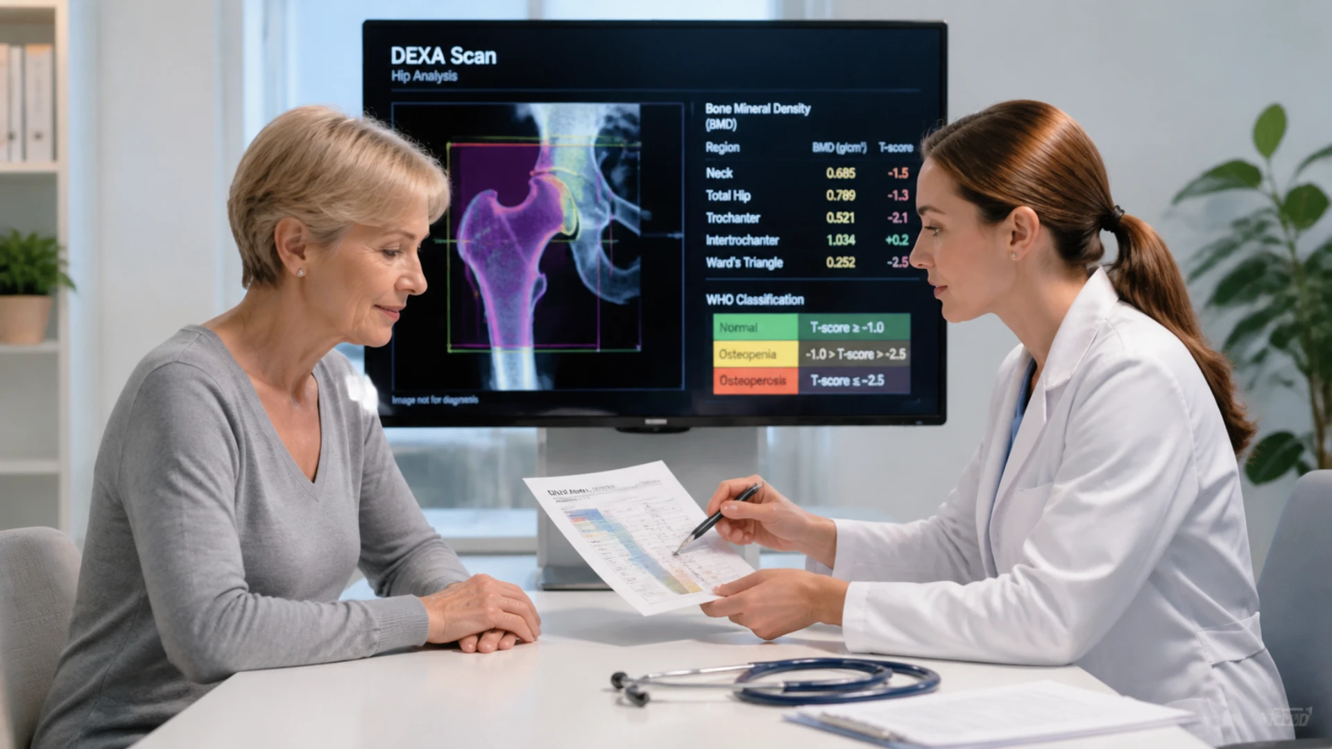
A DEXA (dual-energy X-ray absorptiometry) scan works by directing two low-dose X-ray beams through your body — typically at the hip and lower spine. The denser your bone, the more of that beam it absorbs, and the higher your bone mineral density (BMD) score. It's a useful, widely available, low-radiation tool.
But there's an important limitation that rarely gets explained in the exam room: a DEXA scan can only resolve two tissue types at a time — bone and soft tissue. Because your body also contains fat, muscle, and bone marrow all layered together, the scanner relies on a mathematical algorithm to estimate what it cannot directly see. Researchers have noted that this means your T-score is not a direct measurement — it is a calculated approximation, and the assumptions built into that algorithm can introduce meaningful error. Studies have found that the inherent measurement uncertainty in a DEXA-derived T-score can be as large as one full T-score point — the precise difference between an “osteopenia” and an “osteoporosis” diagnosis.
Perhaps most striking is this landmark finding: in a study of 8,065 women, 54% of those who experienced a hip fracture did not have osteoporosis according to their DEXA scan. More than half. A broader analysis confirmed that the majority of fractures in the general population occur in women with normal or osteopenic bone density — not clinical osteoporosis. This doesn't mean DEXA scans are useless — they provide genuinely meaningful information. But they are a starting point, not the final word.
Yoga for Osteoporosis: Why Bone Quality Matters as Much as Bone Density
Understanding the Trabecular Bone Score (TBS)
One of the most actionable advances in yoga for bone health and osteoporosis care is also one of the least discussed: the Trabecular Bone Score, or TBS. To understand why it matters, consider the difference between bone density and bone quality.
Bone density tells you how much mineral material is packed into a given volume of bone. Bone quality is a broader concept — it describes the internal architecture of your bone: how it's organized, how efficiently it distributes load, and how well it resists cracking under real-world stress. Think of the difference between a brick wall and a Roman arch. Both can use the same amount of material, but the arch — because of how it's structured — bears far greater weight.
Inside your bones is a spongy inner network called trabecular bone — a honeycomb-like lattice of tiny plates and struts. When this lattice is dense, well-connected, and evenly organized, your bone is resilient. When struts thin out or develop gaps, your bone becomes fragile, even if the overall density reading looks acceptable. TBS analyzes the texture of your existing DEXA scan image to infer this internal microarchitecture — no extra scan, no extra radiation, often no additional cost.
The clinical impact is significant: two women can have identical T-scores yet very different fracture risks, because one has beautifully organized internal bone architecture and the other's has deteriorated. A 2023 study found that adding TBS to standard DEXA results changed the treatment recommendation for approximately 40% of patients — meaning a substantial number of women were either being over-treated or under-treated based on their T-score alone. If your imaging center offers TBS analysis, it is absolutely worth requesting.
Osteoporosis Exercises and Bone Turnover: The Real-Time Picture
What Bone Turnover Markers Tell You
There's a question your DEXA scan fundamentally cannot answer: Is your bone getting stronger or weaker right now, today?
A DEXA scan is a snapshot in time. But your bones are in constant, dynamic motion — old bone being broken down by osteoclasts, new bone being built by osteoblasts. The balance between these two processes matters enormously for bone loss prevention yoga and any other intervention you're undertaking.
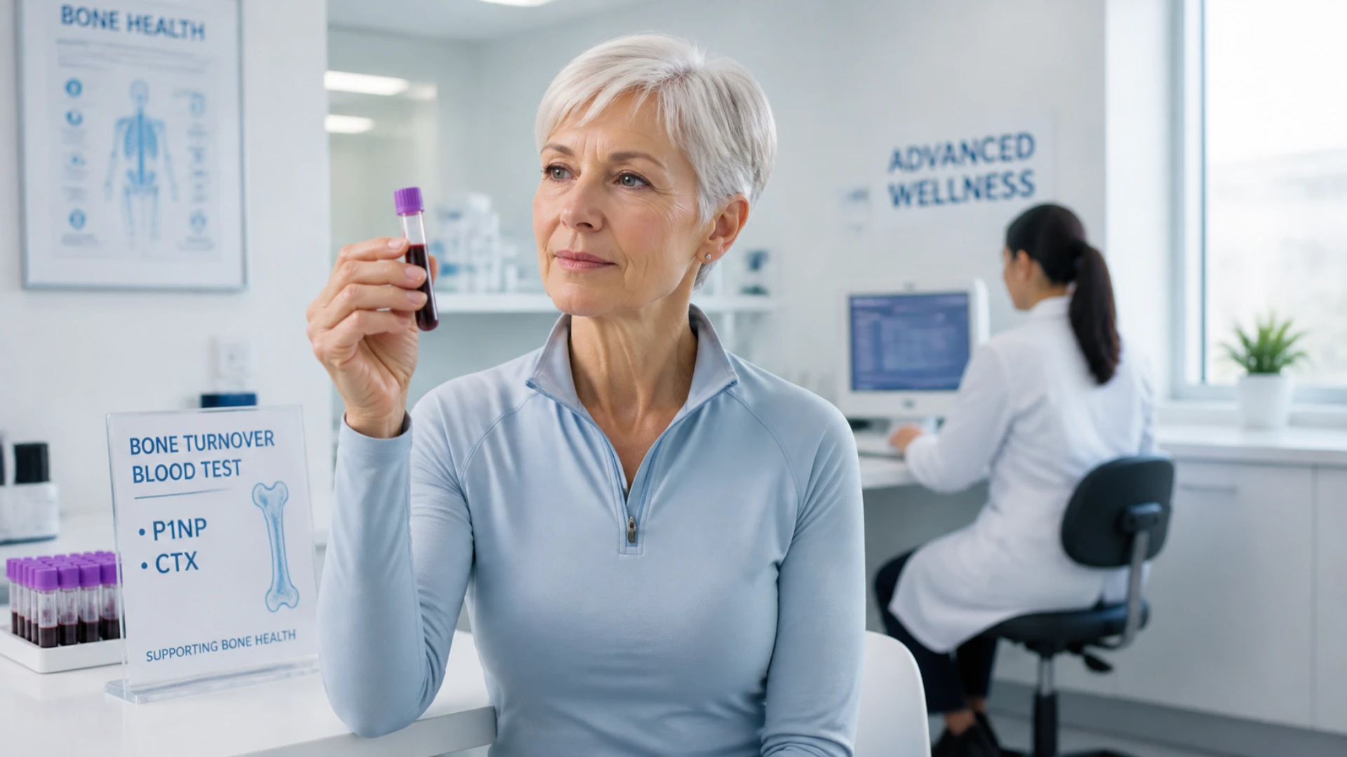
Bone turnover markers are simple blood tests that reveal this balance in real time. The two most important are P1NP (a marker of bone formation) and CTX (a marker of bone breakdown). A 2025 study found that elevated P1NP levels were linked to nearly a 5-fold increase in 10-year fracture risk in postmenopausal women — striking information that no scan can reveal.
These markers are also invaluable for tracking whether your osteoporosis exercises, yoga practice, or medication regimen is actually working — often within weeks, long before a follow-up DEXA scan would show any measurable change. They're standard blood tests your doctor can order at a routine appointment.
Beyond Density: Emerging Tools for Yoga and Aging Well
REMS — Radiation-Free Bone Assessment
For women who want additional information beyond what a DEXA provides — or whose DEXA results may be distorted by spinal arthritis, scoliosis, or other structural changes — a newer tool called REMS (Radiofrequency Echographic Multi-Spectrometry) offers a compelling alternative.
REMS uses ultrasound rather than X-rays to assess bone at the lumbar spine and femoral neck. Because it uses sound waves traveling through bone in multiple planes, it is far less susceptible to the distortions that commonly affect DEXA results in women over 50. Arthritic changes in the spine, for example, can cause DEXA scans to overestimate bone density — registering calcification as apparent bone mass. REMS is unaffected by this.
Crucially, in addition to providing a T-score, REMS produces a fragility score — an assessment of how likely your bone actually is to fracture under real-world stress. One specialist shared the case of a 63-year-old, physically active physician whose DEXA T-score of -2.6 placed him firmly in the osteoporosis range, prompting doctors to tell him to stop skiing. His REMS scan returned a T-score of -1.5, a fragility score in the normal range, and a 10-year fracture risk recalculated at 1–2%. The honest caveat: REMS is not yet a standard clinical replacement for DEXA — most physicians will not prescribe treatment based on REMS results alone — but it can add a meaningful new perspective, particularly when DEXA results seem inconsistent with your functional physical capacity.
HR-pQCT — The 3D Blueprint of Your Bone
At the research frontier sits HR-pQCT (high-resolution peripheral quantitative CT) — a technology that creates a full three-dimensional map of your bone architecture, showing not just density but the precise structural organization layer by layer. Think of it as the difference between knowing how heavy a bridge is versus seeing its full engineering blueprint.
A major multi-site prospective study found that HR-pQCT-based bone strength measures predicted fractures significantly better than standard DEXA — and identified women at high fracture risk who would have been classified as low-risk by bone density alone. Even women with normal bone density who went on to fracture showed significant impairment in bone microarchitecture compared to those who didn't. HR-pQCT remains largely a research tool for now, but the science behind it is actively reshaping how clinicians think about strengthen bones with yoga and other movement-based interventions.
You Are Not Fragile — You Are Trainable
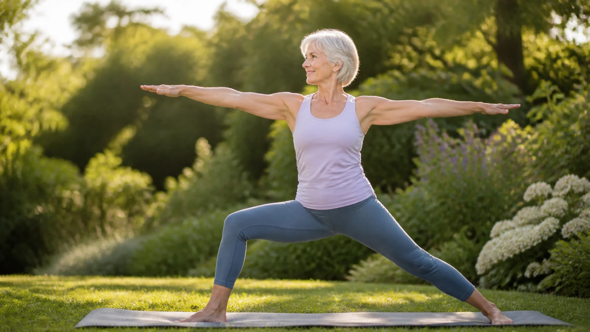
Here is the most important thing to understand from all of this.
A DEXA scan is a two-dimensional, algorithmically derived estimate of bone mineral density at two sites in your body. It says nothing about the quality of your bone architecture, the metabolic activity inside your bones right now, the strength of the muscles that absorb impact, the agility of your nervous system, or your ability to catch yourself mid-stumble. And more than half of women who experience a hip fracture don't even have osteoporosis by DEXA criteria.
Fracture risk is complex, multifactorial, and deeply shaped by factors you can actively work on. Bone density yoga and targeted movement practices address the structural and functional dimensions that scans simply don't capture: muscle strength, balance, proprioception, coordination, and the neuromuscular resilience that protects you when the unexpected happens. The women who fare best as they age are not those who restrict themselves out of fear — they are the ones who keep moving intelligently, building the physical reserves that protect bones in ways no T-score ever could.
Your T-score is one data point. It is not a verdict. And it is certainly not the measure of your vitality or your future.
Your Next Step
Understanding yoga for bone health means understanding the whole picture — bone quality, bone turnover, balance, strength, and resilience. If you're ready to move beyond the T-score and start building the kind of full-body protection that research shows actually matters, there's a beautiful place to start.
Dr. Loren Fishman and Ellen Saltonstall — two of the world's foremost experts on yoga poses for osteoporosis — have developed a free, research-informed introductory series designed specifically for adults navigating osteoporosis and osteopenia. It's safe, evidence-based, and grounded in the same science discussed in this article.
👉 ACCESS THE FREE 3-PART YOGA FOR OSTEOPOROSIS SERIES AT YOGAUONLINE →
References:
-
Järvinen TLN, Michaëlsson K, Aspenberg P, Sievänen H. “Osteoporosis: the emperor has no clothes.” J Intern Med. 2015;277(6):662–673. https://pubmed.ncbi.nlm.nih.gov/25832448/
-
Wainwright SA et al. “Hip Fracture in Women without Osteoporosis.” J Clin Endocrinol Metab. 2005;90(5):2787–2793. https://academic.oup.com/jcem/article/90/5/2787/2836844
-
Yoo JI et al. “Two-Thirds of All Fractures Are Not Attributable to Osteoporosis and Advancing Age.” J Clin Endocrinol Metab. 2019;104(8):3514–3520. https://academic.oup.com/jcem/article/104/8/3514/5427152
-
Pasco JA et al. “The population burden of fractures originates in women with osteopenia, not osteoporosis.” Osteoporos Int. 2006;17(9):1404–1409. https://pubmed.ncbi.nlm.nih.gov/16699736/
-
Martineau P, Silva BC, Leslie WD. “Utility of trabecular bone score in the evaluation of osteoporosis.” Curr Opin Endocrinol Diabetes Obes. 2017;24(6):402–410. https://pubmed.ncbi.nlm.nih.gov/28863001/
-
Samelson EJ et al. “Cortical and trabecular bone microarchitecture as an independent predictor of incident fracture risk.” Lancet Diabetes Endocrinol. 2019;7(1):34–43. https://pubmed.ncbi.nlm.nih.gov/30503163/
-
Johansson H et al. “A meta-analysis of reference markers of bone turnover for prediction of fracture.” Calcif Tissue Int. 2014;94(5):560–567. https://pubmed.ncbi.nlm.nih.gov/24590144/
-
Ensrud KE et al. “A comparison of prediction models for fractures in older women: is more better?” Arch Intern Med. 2009;169(22):2087–2094. https://pubmed.ncbi.nlm.nih.gov/20008691/
-
Gonnelli S, Caffarelli C, Nuti R. “The utility of ultrasonometry in the management of osteoporosis.” Aging Clin Exp Res. 2013;25(5):503–511. https://pubmed.ncbi.nlm.nih.gov/24046064/